Le sildénafil présent dans Kamagra exerce une inhibition réversible de la PDE5, modulant la cascade GMPc et favorisant une vasodilatation localisée. L’absorption digestive varie selon la forme utilisée, comprimés classiques ou gels oraux. La distribution tissulaire est large et la liaison protéique élevée, avoisinant 96 %. La métabolisation hépatique génère un métabolite actif contribuant à l’effet pharmacologique global. La demi-vie reste courte, avec disparition plasmatique en quelques heures. Les interactions significatives concernent surtout les nitrés organiques et inhibiteurs puissants du CYP3A4. Dans les publications techniques, kamagra en ligne est souvent cité dans le cadre d’analyses comparatives portant sur les différences de formulations et de cinétique d’absorption.
Microsoft word - 14_swamy.doc
ɀ ɍ Ɋ ɇ Ⱥ Ʌ ɋ Ɍ Ɋ ɍ Ʉ Ɍ ɍ Ɋ ɇ Ɉ Ƀ ɏ ɂ Ɇ ɂ ɂ
CRYSTAL STRUCTURE OF LANSOPRAZOLE SULFONE
2007 G . Y . S . K . S w a m y * , K . R a v i k u m a r Laboratory of X-ray Crystallography, Indian Institute of Chemical Technology, Hyderabad 500 007, India
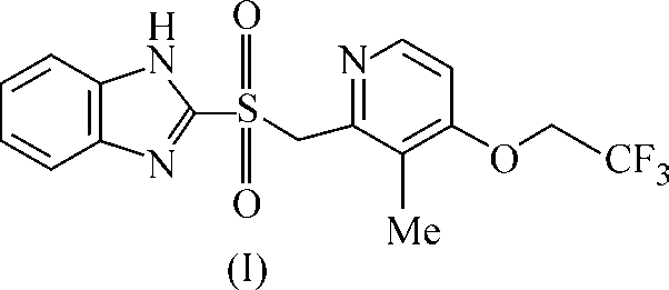
The structure of 2-({[ 3-methyl-4-(2,2,2-trifluoroethoxy)-2-pyridinyl]methyl}sulfonyl)-1H-1,3-benzimidazole, C16H14N3O3F3S, has been solved. The compound belongs to a monoclinicspace group (P 21/c) with cell parameters a = 8.8693(9), b = 23.369(2), c = 8.6141(8) Å,E = 104.68(1)q, V = 1727.2(3) Å3, Z = 4. The final R and wR(F 2) values were 0.070 and 0.147,respectively. The title molecule has a cZc shape in the crystal structure. The fused benzimidaz-ole moiety and the pyridine ring are nearly coplanar. The molecules are linked by N—H…N
and C—H…O hydrogen bonds into chains of edge-fused R 2 (14), R 2 (8), and R 2 (18) rings
along the c-axis. The crystal lattice is further strengthened by S—S-stacking interactions. K e y w o r d s: Crystal structure, benzimidazole, X-ray diffraction, hydrogen bonding. Introduction. The benzimidazole (BI) nucleus is an essential part of many medicinally useful
drugs such as omeprazole, lansoprazole, astemizole and emedastine [ 1, 2 ]. Further, derivatives of BIhave proved to be of considerable value as anthelmintic and antineoplastic [ 3 ], antibacterial and anti-fungal [ 4 ] agents. Such compounds are increasingly being studied in the context of modeling biologi-cal systems [ 5, 6 ]. In continuation of our work [ 7 ] on such compounds, we report here the crystalstructure of the title compound (I). Experimental. The title compound was dissolved in methanol and suitable crystals of X-ray
quality were obtained by slow evaporation.
Diffraction data were measured at room temperature with a Bruker SMART CCD area detector
[ 8 ]. Preliminary lattice parameters and orientation matrix were obtained from three sets of frames. Intensity data were collected using graphite-monochromated MoKD radiation (O = 0.71073 Å).
Integration and scaling of intensity data were accomplished using SAINT [ 8 ]. The structure was
solved by direct methods and refined by a full matrix least-squares procedure based on F 2 [ 9 ]. Non-hydrogen atoms were refined with anisotropic displacement parameters and hydrogen atoms were in-cluded in calculated positions in the riding model approximation. The details of the data collection andrefinement are summarized in Table 1. The geometry and molecular graphics were computed usingprograms PARST [ 10 ], ORTEP-3 [ 11 ], and PLATON [ 12 ]. Selected bond lengths and angles arelisted in Table 2. Results and Discussion. The title molecule has cZc shaped conformation. The stock of cZc com-
prises S1—C8 bond. The left arm (top) and the right arm (bottom) are composed of fused BI system
Crystal data and experimental details
Observed data (I > 2V(I ))
Selected bond distances d (Å) and angles M (deg.)
and trifluoroethoxy-2-pyridinyl moiety (Fig. 1). The BI moiety is planar, within maximum deviationof 0.006(2) Å for the N1 atom. The C—N bond lengths of the imidazole ring are in the range 1.300—1.398(4) Å that is shorter than the single bond length of 1.48 Å and longer than the typical C=Ndistance of 1.28 Å, indicating partial double bond character. The observed lengths can be understoodas a result of conjugation in the heterocycle. Further, the exocyclic angles around atom N2 showconsiderable asymmetry, although the sum of the valent angles around N2 is 360.0q, indicating nosignificant pyramidalization of this atom. The title molecule (I) adopts a nearly trans conformation(C7—S1—C8—C9 angle of 156.2(3)q) which is close to the conformation of omeprazole (179q) [ 13 ]but in contrast with a conformation found in lansoprazole (–96.0(2)q) [ 14 ]. The pyridine ring isalmost coplanar with the BI ring system, with the dihedral angle of 8.1(1)q between their least-squaresplanes. The plane containing trifluoroethoxy group makes angle of 11.0(2)q with the pyridine ring. The dihedral angle between the sulfonyl moiety and the BI ring system is 54.9(1)q. Fig. 1. ORTEP drawing of (I) showingthe atom numbering scheme. Thermalellipsoids are drawn at 30 % probabi- lity level
Fig. 2. Fragment of the crystal struc-ture of (I) showing the formation of an
and R2 (28) rings along the c-axis
The sulfonyl oxygen O2 is involved in hydrogen bonding interactions with two types of atoms
(C8—H8B…O2 and C14—H14A…O2). While the first interaction generates R 2 (8) type rings, the
second one produces R 2 (18) rings. Inversion-related pairs of molecules (pseudo dimers) are joined
Hydrogen bonding geometry (Å, deg.)
R 2 (8) edge-fused rings (Fig. 2) [ 15 ].
the fluorine atoms (F1), C(15) type chains
being thus formed along the [001] direc-tion. These chains are further connectedto the neighboring chains via C—H…N Symmetry codes: (i) x+1, y, z+2; (ii) –x+1, –y+1, –z+1; (iii) –x,(C(10) type, Table 3) hydrogen bonds.
–y+1, –z; (iv) x, y, z–1.
Stacking interactions between the pyridine and the imidazole ring of the BI moiety with a spacing
of 3.5 Å provide further stability to the crystal lattice. Acknowledgements. The authors thank M/s Lee Pharma for help in providing the compound and
Dr. J.S. Yadav, Director, IICT, Hyderabad, for his kind encouragement.
1. Sakai T., Hamada T., Awata N., Watanabe J. // J. Pharmacobio. Dyn. – 1989. – 12. – P. 530 – 536. 2. Nishina K., Mikawa K., Maekawa N. et al. // Anesth. Analg. – 1996. – 82. – P. 832 – 836. 3. Ram S., Wise D.S., Wotring L.L. et al. // J. Med. Chem. – 1992. – 35. – P. 539 – 547. 4. Kus C., Goker H., Ayhan G., Ertan R. // Farmaco. – 1996. – 51. – P. 413 – 417. 5. Bouwman E., Driessen W.L., Reedijk J. // Coord. Chem. Rev. – 1990. – 104. – P. 143 – 172. 6. Pujar M.A., Bharamgoudar T.D., Satyanarayana D.N. // Transition Met. Chem. – 1988. – 13. – P. 423 –
7. Swamy G.Y.S.K., Ravikumar K. // Acta Crystallogr. – 2005. – E61. – P. o4200 – o4202. 8. SMART & SAINT. Software reference manuals. Versions 6.28a & 5.625. – Bruker Analytical X-ray Sys-
tems Inc., Madison, Wisconsin, U.S.A., 2001.
9. Sheldrick G.M. SHELXS-97 and SHELXL-97. Programs for crystal structure solution and refinement. –
University of Gottingen: Germany, 1997.
10. Nardelli M.J. // J. Appl. Crystallogr. – 1995. – 28. – P. 659. 11. Farrugia L.J. // Ibid. – 1997. – 30. – P. 565. 12. Spek A.L. // Ibid. – 2003. – 36. – P. 7 – 13. 13. Ohishi H., In Y., Ishida T. et al. // Acta Crystallogr. – 1989. – C45. – P. 1921 – 1923. 14. Vyas K., Sivalakshmidevi A., Om Reddy G. // Ibid. – 2000. – C56. – P. e572 – e573. 15. Bernstein J., Davis R.E., Shimoni L., Chang N.L. // Angew. Chem. Int. Ed. Engl. – 1995. – 34. – P. 1555 –
Periodico missionario allegato ANNO 2 . NUMERO 3 . FEBBRAIO2010 al “Sette Giorni Comunità” > EDITORIALE A Milano è stato ammazzato un ragazzo parlano e sparlano crediamo si nasconda il e clarinettista dell’Orchestra di Via Padova, desiderio di espellere gli immigrati. Tutti gli rilasciata al free press “MetroMilano”. immigrati. Corradi: «Tutti pensano che il
Mædica - a Journal of Clinical Medicine Update in Diabetology Cristian GUJA, MD, PhDa; Radu LICHIARDOPOL, MD, PhDa,baThe National Institute of Diabetes, Nutrition and Metabolic Diseases „Prof. NC bThe University of Medicine and Pharmacy “Carol Davila”, Bucharest, Romania S everal important developments in the does not decrease the frequency of cardiovas-diagnosis and treatment o
 ɀ ɍ Ɋ ɇ Ⱥ Ʌ ɋ Ɍ Ɋ ɍ Ʉ Ɍ ɍ Ɋ ɇ Ɉ Ƀ ɏ ɂ Ɇ ɂ ɂ
CRYSTAL STRUCTURE OF LANSOPRAZOLE SULFONE
ɀ ɍ Ɋ ɇ Ⱥ Ʌ ɋ Ɍ Ɋ ɍ Ʉ Ɍ ɍ Ɋ ɇ Ɉ Ƀ ɏ ɂ Ɇ ɂ ɂ
CRYSTAL STRUCTURE OF LANSOPRAZOLE SULFONE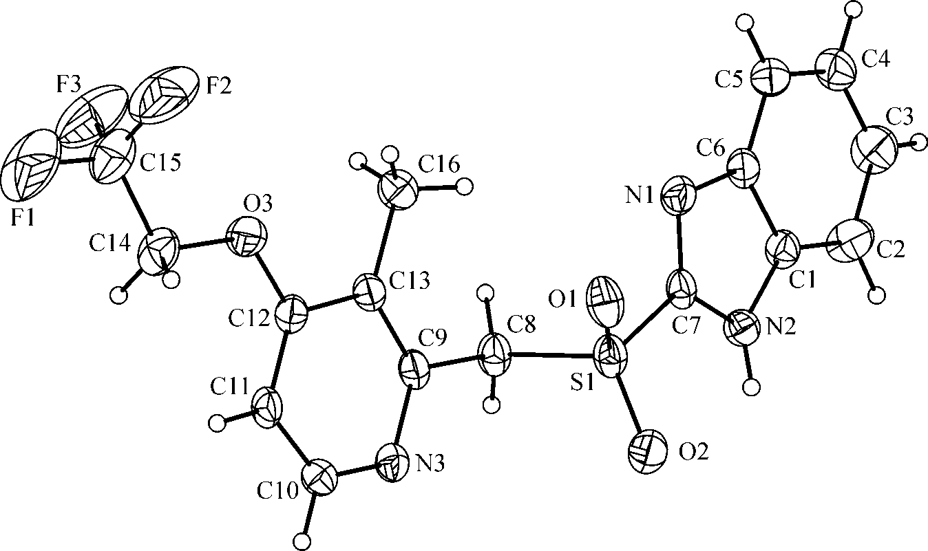
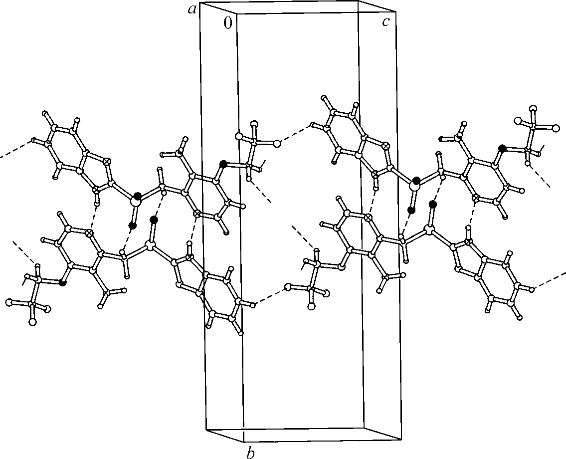 Fig. 1. ORTEP drawing of (I) showingthe atom numbering scheme. Thermalellipsoids are drawn at 30 % probabi- lity level
Fig. 2. Fragment of the crystal struc-ture of (I) showing the formation of an
and R2 (28) rings along the c-axis
The sulfonyl oxygen O2 is involved in hydrogen bonding interactions with two types of atoms
(C8—H8B…O2 and C14—H14A…O2). While the first interaction generates R 2 (8) type rings, the
second one produces R 2 (18) rings. Inversion-related pairs of molecules (pseudo dimers) are joined
Hydrogen bonding geometry (Å, deg.)
R 2 (8) edge-fused rings (Fig. 2) [ 15 ].
Fig. 1. ORTEP drawing of (I) showingthe atom numbering scheme. Thermalellipsoids are drawn at 30 % probabi- lity level
Fig. 2. Fragment of the crystal struc-ture of (I) showing the formation of an
and R2 (28) rings along the c-axis
The sulfonyl oxygen O2 is involved in hydrogen bonding interactions with two types of atoms
(C8—H8B…O2 and C14—H14A…O2). While the first interaction generates R 2 (8) type rings, the
second one produces R 2 (18) rings. Inversion-related pairs of molecules (pseudo dimers) are joined
Hydrogen bonding geometry (Å, deg.)
R 2 (8) edge-fused rings (Fig. 2) [ 15 ].